BNA members receive international recognition with SfN awards
8th October 2024


20th Nov 2019

Memory is a topic which is of personal interest to us all - why we remember some things better than others, how we can memorise things most easily, what happens in our brain to form memories…
In addition, research into memory is one of the largest areas of research in neuroscience, particularly due to the need to understand and develop treatments for neurological conditions such as epilepsy and Alzheimer’s disease.
Some of the greatest discoveries of the function of different parts of the brain, including those involved in memory, have come through studying those who have neurological conditions as a result of brain injury, both during their lives and by examining their brains after death. One such case, who has been called the most important patient case in the history of brain science, is Henry Gustav Molaison, known to the world as ‘Patient HM’. Suffering from severe epilepsy, in 1953 he underwent major surgery to have a portion of his temporal lobe removed, including parts of the hippocampus and amygdala, from both sides of the brain.
After surgery, it was discovered that Henry was suffering from severe amnesia and although he could remember much of his childhood, he struggled to remember events which occurred during the years leading up to the surgery. He also suffered from anterograde amnesia and was unable to create any new memories. In the many tests and studies that Henry willingly took part in until his death aged 82, evidence was accumulated which allowed a major shift in neuroscientists’ understanding of memory systems. In particular, it lead to a reassessment of the then-accepted notion that memory functions were spread throughout the brain, demonstrating that there are distinct forms of memory which are located in different parts of the brain.
Nowadays, modern imaging techniques such as MRI allow us to study the brains of patients with neurological conditions and of those with head injuries as well as allowing us to gain a greater understanding of the function of healthy brains. Although these tools have been invaluable, animal models are still routinely used as they allow much greater anatomical precision.
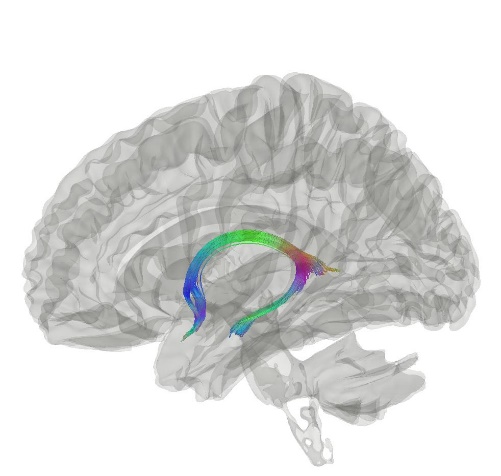 The fornix (meaning ‘arch’ in Latin) is a C-shaped nerve fibre tract linking the hippocampus and diencephalon (and other structures). These two structures are heavily involved in episodic memory, a type of long-term memory that involves conscious recollection of past experiences along with the context in which they occurred – the place where the experience occurred, the time, the emotions that were felt. One example which demonstrates this is the strong link of the disruption of connections between the hippocampal formation (a compound structure in the brain including several brain regions) and the diencephalon (a division of the forebrain, consisting of the hypothalamus and other structures) with anterograde amnesia.
A new study (Mathiasen et al., 2019) available in the BNA’s journal, ‘Brain and Neuroscience Advances’, examined the path that the fibres in the fornix take through the brain beyond the temporal lobe, a pathway which until now has been poorly described. These routes were examined in three species – the mouse, rat and macaque monkey. Although different methods and species were used, the principal pattern which arose was the same.
Despite not being able to trace all fibres to their target areas, new information was obtained as to the routes of many of the fibres which project from the hippocampal formation to the anterior thalamic nuclei and mammillary bodies (part of the diencephalon).
Understanding the path that these fibres take will allow us to better understand the contribution that these different fibres and structures make to memory and cognition.
Regularly, research is done on subjects who have lesions in parts of their brain – so-called disconnection analyses. The present findings will be invaluable in helping to interpret diencephalic disconnection studies in rats as well as the fornix sections studied in patients with epilepsy.
To read the full article, click here.
Mathiasen, M., Louch, R., Nelson, A., Dillingham, C. and Aggleton, J. (2019). Trajectory of hippocampal fibres to the contralateral anterior thalamus and mammillary bodies in rats, mice, and macaque monkeys. Brain and Neuroscience Advances, 3, p.239821281987120.
Image of fornix:
By Yeh, F. C., Panesar, S., Fernandes, D., Meola, A., Yoshino, M., Fernandez-Miranda, J. C., ... & Verstynen, T. (2018). Population-averaged atlas of the macroscale human structural connectome and its network topology. NeuroImage, 178, 57-68. - http://brain.labsolver.org/, CC BY-SA 4.0, https://commons.wikimedia.org/w/index.php?curid=78298712
Information about patient HM:
Myers, J. (2017). Brain case study: Patient HM. [online] Big Picture. Available at: https://bigpictureeducation.com/brain-case-study-patient-hm [Accessed 19 Nov. 2019].