Exploring the World of Neuroscience - by BNA Members
Earlier this year, we invited our BNA Member community to share images capturing their everyday neuroscience - and the results were truly captivating: both visually striking and scientifically insightful.
We received an incredible range of submissions that reflected the diversity of our membership, spanning academia, industry, and clinical practice. From the lab bench to the microscope and patient care, each image offered a unique glimpse into the daily work that drives neuroscience forward.
The judging panel, made up of BNA Council Members, had a tough task selecting just twelve standout images from the many impressive submissions. These selected images now feature in our 2025/26 academic year calendar and were proudly exhibited at the ‘Inform’ stand during BNA2025.
Delegates at the conference then had the chance to vote for their favourite and pick up a calendar - and we're delighted to reveal the winning image, along with the full set of shortlisted entries.
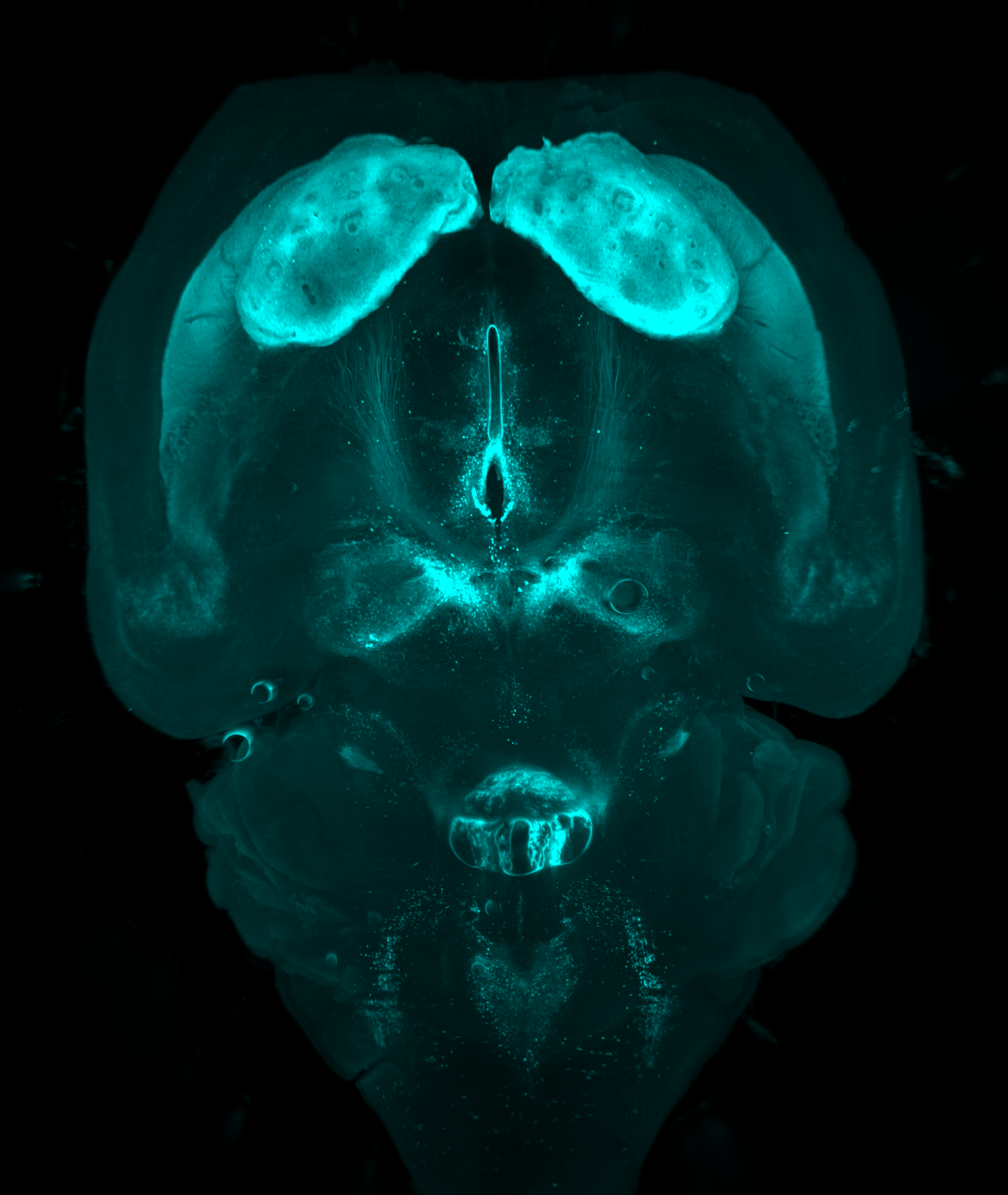
Our winner was an image submitted by Professional Member, Dr Cristina Martinez-Gonzalez.
'A whole mouse brain labelled against tyrosine hydroxylase to visualize dopaminergic, adrenergic and noradrenergic neurons and their projections (cyan). Tissue was cleared using RatDISCO and imaged through light-sheet microscopy.'
On winning the competition Dr Martinez-Gonzalez said: "Thank you to BNA for this fantastic recognition that highlights my passion for microscopy. And thank you for bringing together such a diverse community of scientists and for giving opportunities to show our skills beyond the lab."
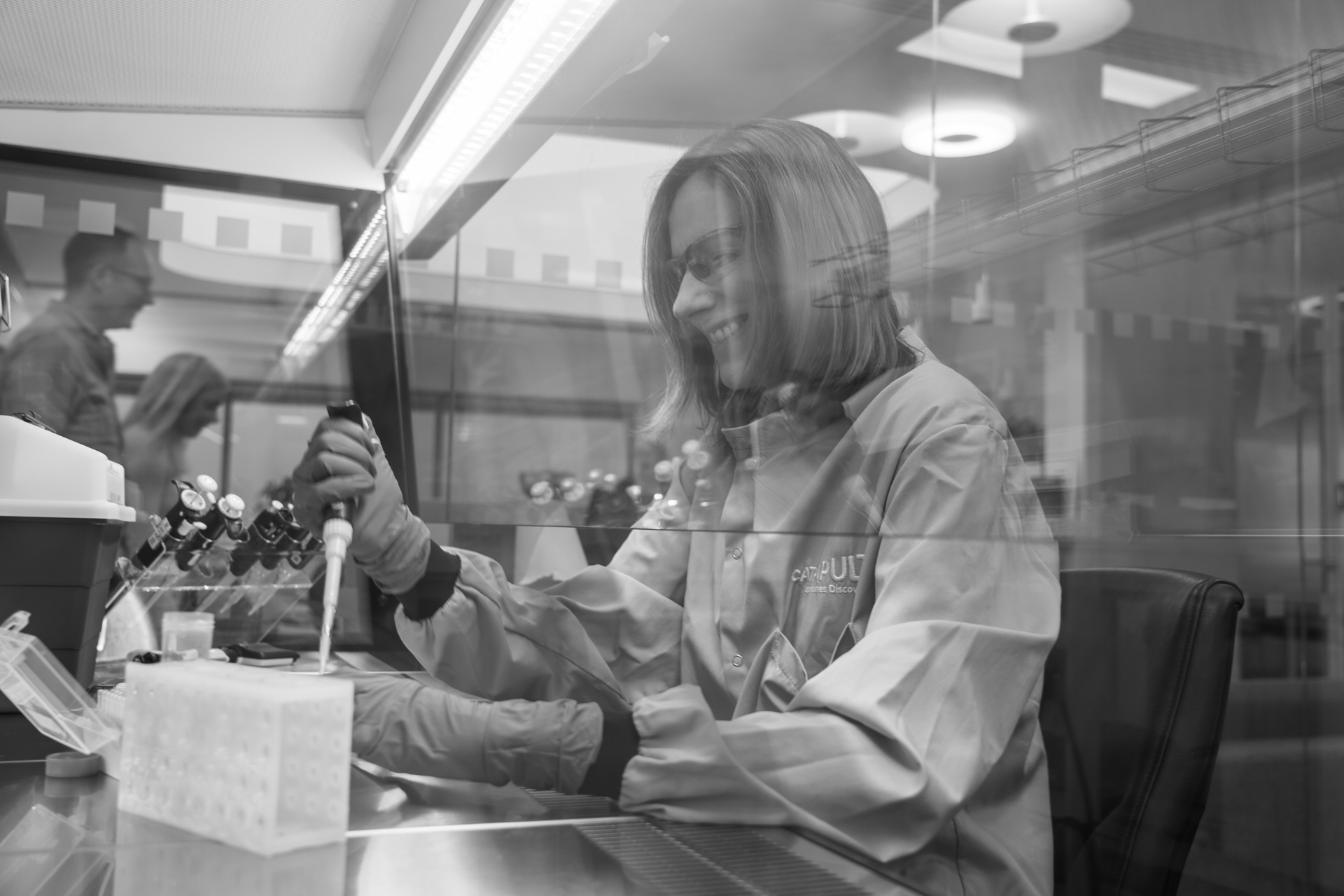
Image by Professional Member, Dr Ekta Patel
'Lead Scientist in Neurobiology at work in the hood.'
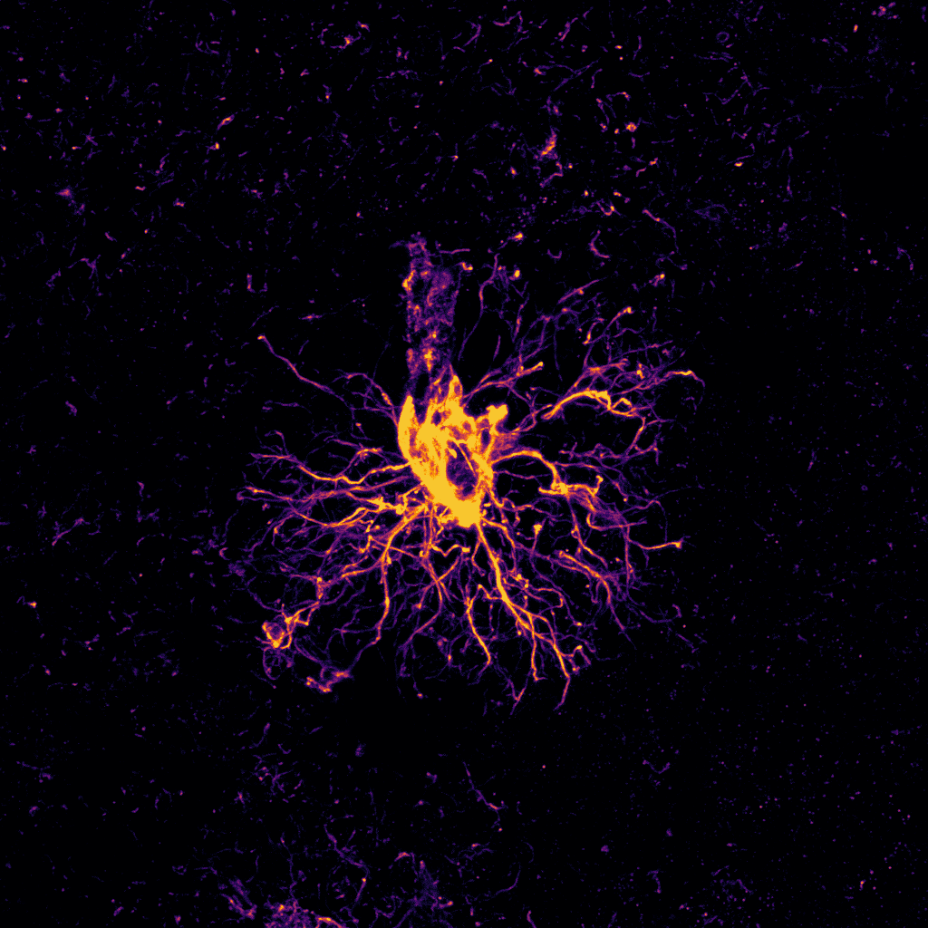
BNA Postgraduate Member (PhD) Kratika Mujmer
'A lone Aldh1l1 positive astrocyte captured using confocal microscopy.'
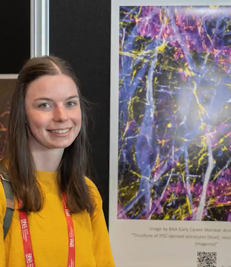
Early Career Member, Dr Anna-Lena Zepernick was pleased to see her image in the shortlisted gallery at BNA2025.
Her image depicts 'Triculture of iPSC-derived astrocytes (blue), neurons (yellow) and microglia (magenta).'

Postgraduate Member, Poppy Firchau
Image 'shows a fly eye being photographed at x8 magnification with a Zeiss 3.0 microscope. Images taken in this way are used to determine a 'rough eye phenotype' which may be present in Alzheimer's Drosophila models due to a loss of retinal cells when proteins are misexpressed with GMR-drivers in a UAS-GAL4 system (a method of gene expression in Drosophila using yeast transcription factors).'
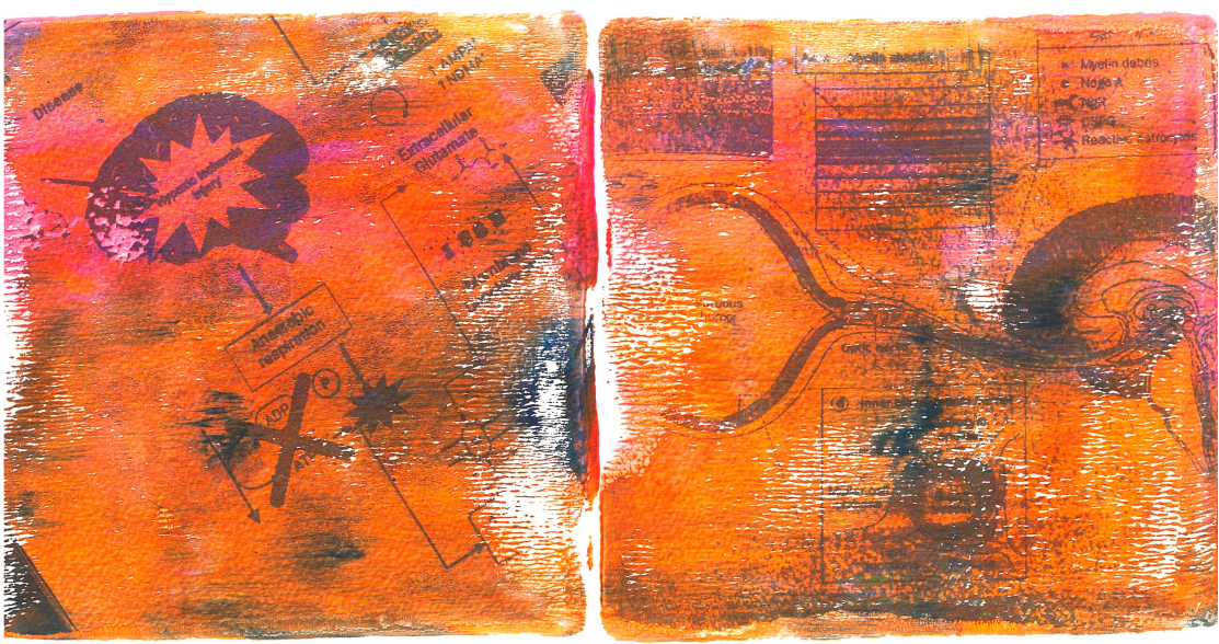
Postgraduate Member (PhD) - Dipa Begum
'Using the tactile Gelli printing art technique, this piece showcases two avenues of neuroscience research from two PhD students at the Perinatal Brain Repair group at UCL. At a workshop facilitated by Dr Jennifer Crouch, myself and my lab colleague Gelli printed a figure which best represented our research focus, one therapeutic and one diagnostic. Despite our different approaches, our work coalesces to improve and advance perinatal brain repair research.'
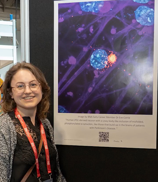
Early Career Member, Dr Eve Corrie was pleased to see her shortlisted image exhibited in the ‘Inform’ gallery at BNA2025.
'This is a human iPSC-derived neuron with a Lewy body-like inclusion of misfolded, phosphorylated α-synuclein, like those that build up in the brains of patients with Parkinson's Disease.'
BNA Chief Executive, Laura Ajram says:
“This competition has been a wonderful celebration of the creativity, passion, and diversity within our BNA member community. The images submitted go beyond data - they tell powerful stories about the everyday work that drives neuroscience forward. It’s inspiring to see such a range of perspectives, from early-career researchers to seasoned professionals, across academia, industry, and clinical practice. We’re proud to showcase this work and the people behind it.”
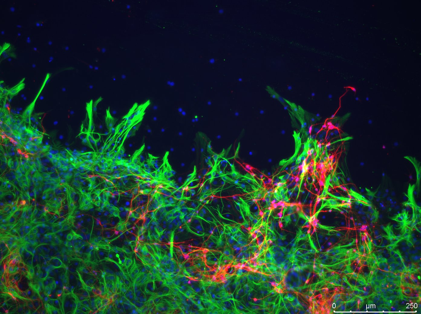
BNA Postgraduate Member (PhD) Zoe Dombros-Ryan
'Neurons (TUJ1/red) and astrocytes (GFAP/green) dancing along an injury border. Fluorescent image showing how cultured primary mouse cortical neurons and astrocytes behave in the presence of a simulated penetrating traumatic brain injury.'
Professional Member - Dr Mick Craig
'This is from a study exploring the effects of cytokine overexpression in astrocytes. In magenta, there is an astrocyte overexpressing TNFa (reported via TdTomato). Yellow shows an IBA1 immunostain for microglia - the striking feature of this image is that microglia have become reactive in a zone around the astrocyte while those further away remain in a resting state. Cyan shows a nuclear DAPI stain.'
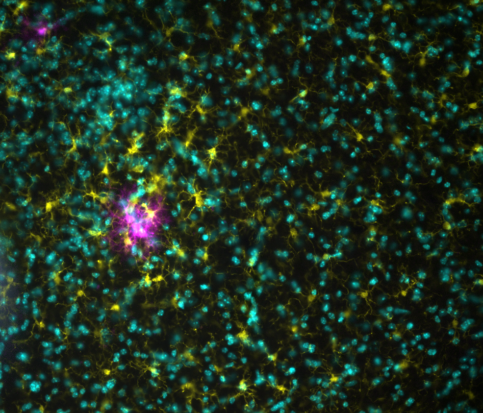
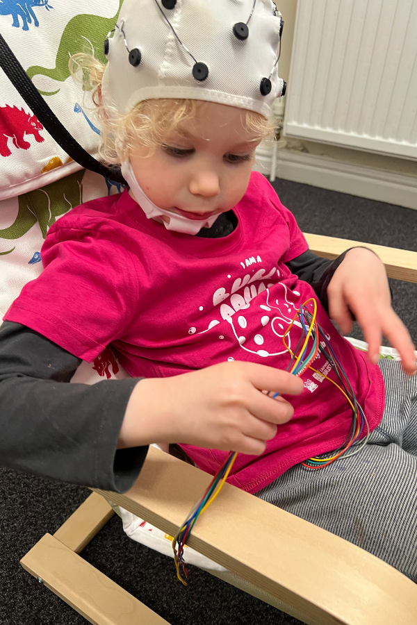
Image by Professional Member, Dr Edwin Dalmaijer
'In my lab, we measure brains and stomachs, and find that disgust changes neurogastric interactions. We're investigating if this underlies fussy eating in children, but sometimes we're interrupted by sibling tantrums!'
Image by Undergraduate Member, Ruxandra Birea
'Astrocytes (green) and Neurons (purple) mimicking aurora borealis, the northern lights. Fluorescence microscopy showing Astrocytes (GFAP, green) and Neurons (TUJ1, purple), aligned along parallel polymer nanofibers, going in a clear direction. Nuclei shown in blue (DAPI). This directed neural growth could help reconnect damaged spinal cord and promote regeneration.'
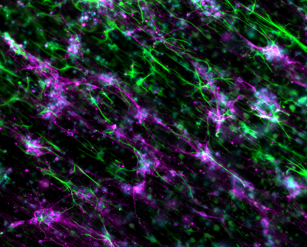
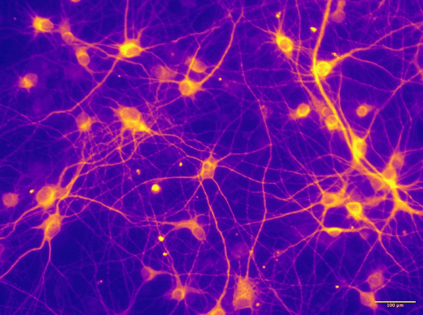
Image by Postgraduate Member, Aparna Maruvada
'Brain Tapestry - A glowing galaxy of thoughts.'
We hope these images showcase the many facets of neuroscience - from the people and settings to the stunning beauty revealed through the microscope - and inspire the next generation of researchers, clinicians, and innovators. Neuroscience is beautiful, and after such an incredible response, we just might make this an annual tradition!